Do I need an Xray or a Scan?
When to x-ray, MRI or ultrasound for foot Injuries? This is a question we hear often at Waikato Podiatry, and here are some guides to help you better understand the role of imaging in podiatry and when it can help.
If you require an X-ray, MRI, or ultrasound, we can prescribe and organise this for you at Waikato Podiatry Clinic and ensure you get the right one for your problem. This will enable us to diagnose your condition and create a treatment plan.
Lets look at the different types of imaging and what they can and cannot offer.
X-Ray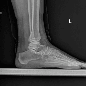
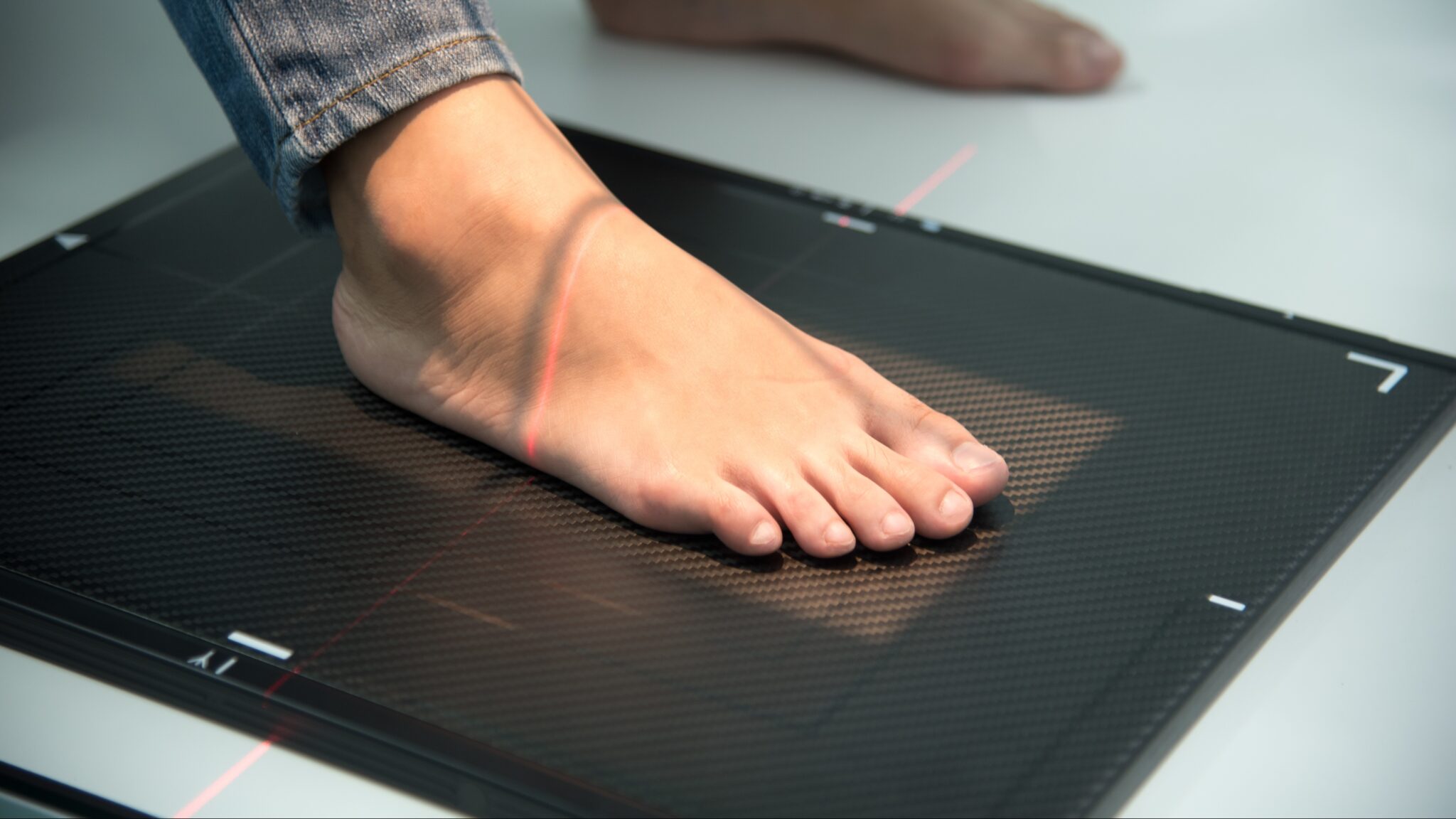
X-Ray is the baseline level of imaging and it is used to assess bones and joints. It can pick up and identify fractures, arthritic changes and some alignment issues. It is not good at picking up stress fractures or soft tissue injuries. In the Podiatry context X-rays should always be done if possible, in a weight bearing or standing situation. This allows a better interpretation of joints of the foot in a more real-life application.
Ultrasound
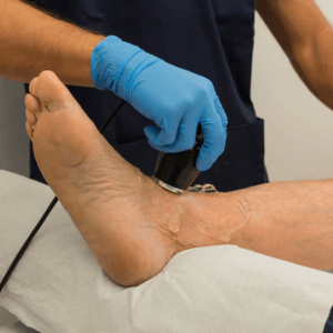
Ultrasound uses soundwaves to bounce off the soft tissue, it is used for examining ligaments tendons and other soft tissue structures. It is able to identify masses and in some instances damage to soft tissue. A problem with ultrasound is that it is very user dependent and it is not easy to interpret the subtle differences between some conditions; for example, some masses cannot be differentiated. It is relatively easy to access and a cheap modality that is useful for the clinician, some clinicians have immediate access to these in their rooms.
MRI, Magnetic Resonance Imaging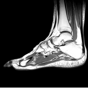
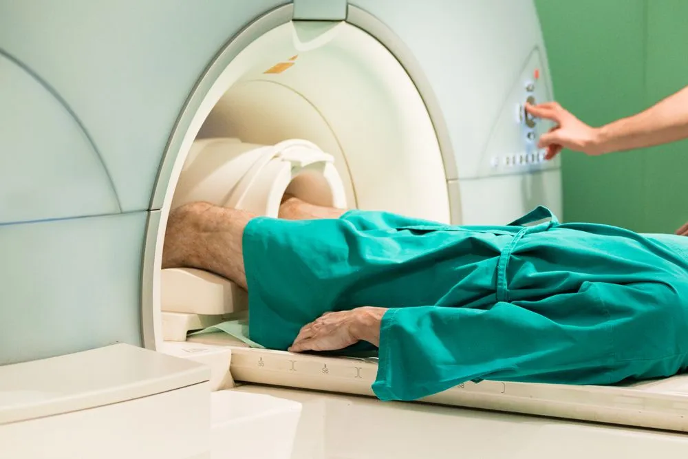
Magnetic Resonance Imagine, MRI is the more complex and detailed imaging modality that has a high sensitivity and specificity to identify damage and changes in bone and soft tissue structures. It is a lot more expensive and therefore harder to access as there are not as many areas that do the scans.
Once again, interpretation of the findings is key in it's use.
Integrating Medical Imaging into Treatment
Medical imaging has become steadily more available over the last 50 years. As with a lot of medical advancements the real benefit lies in how they are used and applied. It is a considered decision of when to x-ray, MRI or ultrasound for foot injuries. All medical images need to be viewed by a skilled clinican to inform the diagnose and treatment plan.
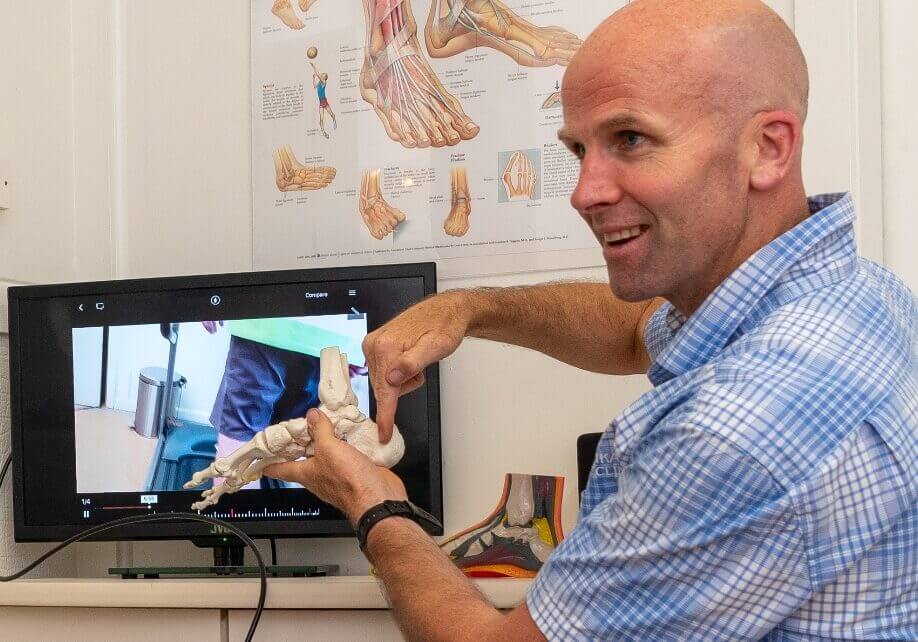
There is a saying "treat the patient, not the X-ray".
This speaks to the fact that imaging will pick up unrelated issues that may be a distraction or a further complication and should not be overly focused on, if they are not relevant or helpful. This is one of the biggest downsides of advanced imaging. There is a lot of research to show that you cannot and must not diagnose exlusively on imaging alone!!
It must always be applied in the clinical context, the pain patterns and presentation is consistent with what is in the scan.
Imaging Guidelines
Here are some guidelines around what conditions we would order investigations for;
- To identify the injured tissue and confirm a diagnosis or give additional clinical information.
- To give an idea of how bad the injury may be and therefore potential healing times.
- To try and narrow the cause of the pain, when it could be a number of things.
- To investigate the position of the bones and other tissues.

When to Image or Scan?
As a clinician, we need to be sure that requesting the imaging is going to add significant value to our treatment plan and therefore your recovery. The vast majoirty of injuries will not require any imaging, as a careful history and clinical decision-making is enough to frame an effective treatment plan. Certain conditions have characteristics which make diagnosis easier.
- Injuries and conditions more likely to require imaging include things such as;
- High force and impact involved
- Severity and mechanism of injury e.g how it happened
- Prolonged intense pain without resolution
- Chronic unresolved problems of an unclear nature.
You may be interested in looking at thge Ottowa ankle rules;
There are an evidence based guise as to when you need an x-ray on your sprained ankle.
Ottawa ankle rules | Radiology Reference Article | Radiopaedia.org
If you do require an x-ray or ultrasound we can organise and prescribe this for you at Waikato Podiatry Clinic and ensure you get the right one for your problem.
One example of requiring  imaging may be falling off a roof and landing on your feet, having significant swelling and intense pain in your heels, and being unable to weight bare on one foot. In this situation, there is a likelihood of significant injury. We would start with an X-ray to check for a fracture, then potentially look at using an ultrasound to check soft tissue damage.

